Forgetting visualized in brain cells
Researchers at the Ludwig-Maximilians-Universität in Munich have achieved a previously unattained quality of visualisation of the brain cells of mice suffering from Alzheimer's disease with the help of X-ray phase-contrast computed tomography.
 here is a growing understanding of what happens in the brain when it is afflicted with Alzheimer's disease. Due to the death of nerve cells, people with Alzheimer's become increasingly forgetful, confused and disoriented. To date, Alzheimer's is incurable. But there are many approaches to getting the disease under control. One of these is an understanding of the neuronal processes that take place in our thinking organ. A team led by Prof. Paola Coan from the Department of Medical Physics and the Department of Radiology at the LMU Hospital in Munich has now gained new findings concerning this process with the help of X-ray phase-contrast computed tomography. Using this imaging technique, the scientists have gained unique insights into the ageing and neurodegeneration of brain cells in mice suffering from Alzheimer's disease.
In western industrial nations, Alzheimer's is one of the most dreaded diseases. At present, it is only possible to slow down the degeneration of the brain cells. Nevertheless, the disease remains incurable. An improved visualization and thus understanding of the mechanisms of Alzheimer's and its processes is key for treatment development. Sophisticated imaging techniques, such as the relatively new X-ray phase-contrast computed tomography, can play a fundamental role. By using this innovative imaging approach, a team led by Prof. Paola Coan from the Department of Medical Physics and the Department of Radiology at the LMU Hospital in Munich has now examined brain cells of mice suffering from Alzheimer's disease and gained new insights into the cellular processes that take place during the course of the disease. Twelve European research institutions were involved in the international project, with researchers from the LMU, the IRCCS Neuromed Institute in Pozzilli (IT) and the Sapienza University in Rome as the leading investigators.
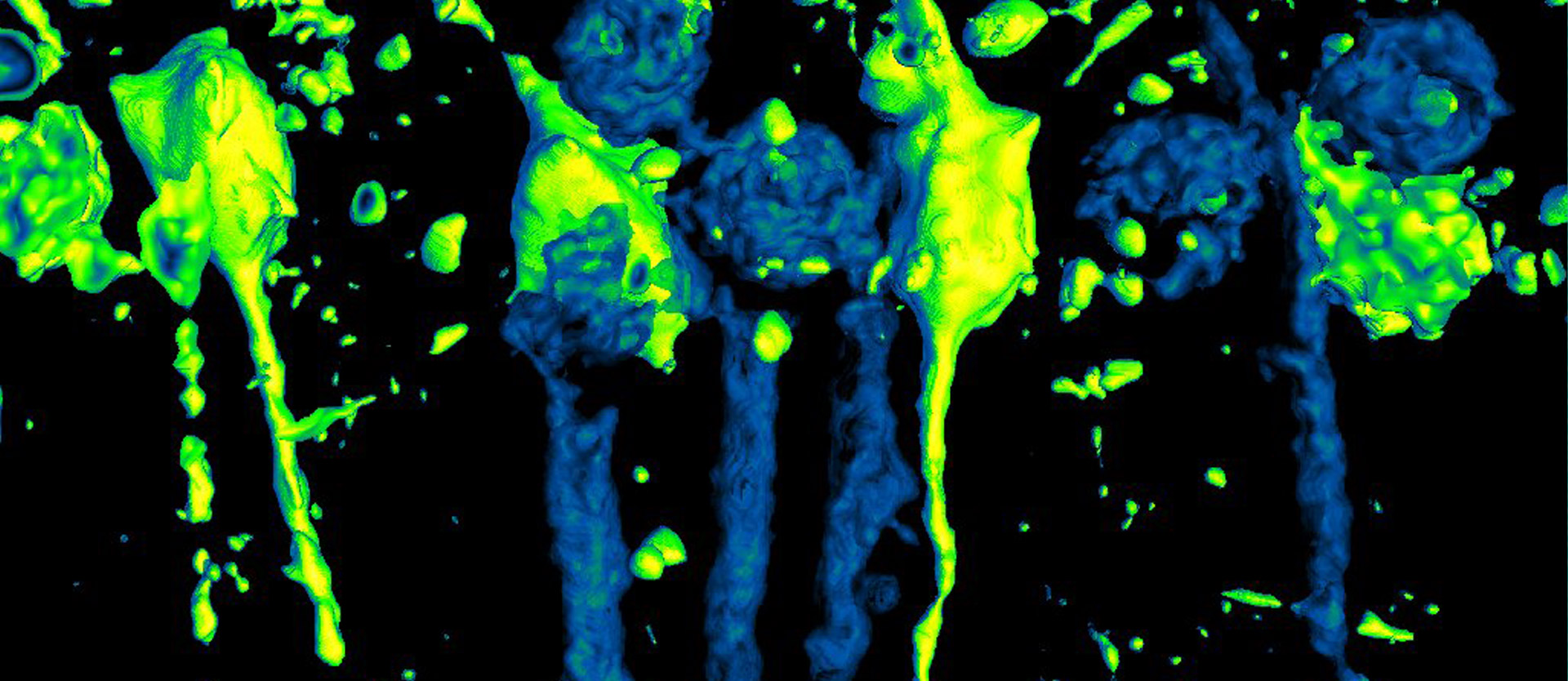
In contrast to conventional X-ray technology, multi-scale X-ray phase-contrast computed tomography opens up the possibility of sensitively imaging not only bones but also X-ray-transparent objects such as soft tissue. The LMU scientists used these 3D imaging techniques to carry out original virtual histological work on small-animal brain tissue, thereby depicting cells affected by Alzheimer's disease in three dimensions and in their almost natural environment. The analysis allowed the team to follow cell ageing and degeneration in detail over time.
"We were able to capture data sets that represent the neuroanatomy of the entire brain," explains Paola Coan. In parallel, the scientists were able to visualize individual cells. "This virtual X-ray phase contrast histological method, which relies on complementary techniques and a multiscale and multimodal approach, allowed us to visualise and quantify cell populations in three dimensions in extended dissected mouse brain samples, as well as to identify diseased neurons by detecting intracellular hyperdensity in the specific brain layers affected in AD," Dr. Giacomo Barbone, first author of this publication, further explains. The data was also correlated with immunohistochemistry results, X-ray nanofluorescence, high-resolution magnetic resonance imaging and transmission electron microscopy results.
With their findings, the researchers have achieved a previously unattainable quality of visualisation of anatomical and pathological three-dimensional brain layers - from the organ level to the intracellular level. "The ability to volumetrically measure extensive populations of degenerating hippocampal cells in deep brain layers without staining or dissection is new and a technical feat," explains Paola Coan.
The new three-dimensional examination method of cells that are in a nearly natural structural state could now be used to test new drug protection strategies that help prevent cells from being affected by Alzheimer's disease.
Original publication:
Giacomo E. Barbone, Alberto Bravin, Alberto Mittone, Alexandra Pacureanu, Giada Mascio, Paola Di Pietro, Markus J. Kraiger, Marina Eckermann, Mariele Romano, Martin Hrabě de Angelis, Peter Cloetens, Valeria Bruno, Giuseppe Battaglia & Paola Coan
X-ray multiscale 3D neuroimaging to quantify cellular aging and neurodegeneration postmortem in a model of Alzheimer’s disease
European Journal of Nuclear Medicine and Molecular Imaging July 19th 2022
DOI: doi.org/10.1007/s00259-022-05896-5
For more information, please contact:
Prof. Dr. Paola Coan
LMU - Lehrstuhl für Experimentelle Physik - Medizinische Physik
Department für Klinische Radiologie der LMU
Am Coulombwall 1, 85748 Garching,
Tel.: 0049 89 289 14062
E-mail: Paola.Coan@physik.uni-muenchen.de
www.lmu-klinikum.de/radiologie/forschung/x-ray-diagnostics-a
Welcome to Photonworld – a platform for knowledge, interesting facts and science on the topic of light. Photonworld aims to provide a basic understanding of the physics of light and its countless phenomena. From medicine, to art and physics: all topics are covered.
Photonworld originated as an initiative of the excellence cluster the Munich-Centre for Advanced Photonics (MAP) and the Laboratory for Attosecond Physics (LAP) at the Max Planck Institute of Quantum Optics (MPQ).


The initiator of the project is Prof. Dr. Ferenc Krausz, Chair of Experimental Physics at the LMU and Director of the MPQ.
Photonworld’s Student Zone reports directly out of the PhotonLab, which is supported by the Munich Center for Quantum Science and Technology (MCQST) and the DFG Research Unit FOR 2783. At the PhotonLab, schoolteachers and students can find up-to-date information about experiments and goings-on at the lab, and plan their own visit to the laboratory.